Abstract
The early sign detection of liver lesions plays an extremely important role in preventing, diagnosing, and treating liver diseases. In fact, radiologists mainly consider Hounsfield Units to locate liver lesions. However, most studies focus on the analysis of unenhanced computed tomography images without considering an attenuation difference between Hounsfield Units before and after contrast injection. Therefore, the purpose of this work is to develop an improved method for the automatic detection and classification of common liver lesions based on deep learning techniques and the variations of the Hounsfield Units density on computed tomography scans. We design and implement a multi-phase classification model developed on the Faster Region-based Convolutional Neural Networks (Faster R-CNN), Region-based Fully Convolutional Networks (R-FCN), and Single Shot Detector Networks (SSD) with the transfer learning approach. The model considers the variations of the Hounsfield Unit density on computed tomography scans in four phases before and after contrast injection (plain, arterial, venous, and delay). The experiments are conducted on three common types of liver lesions including liver cysts, hemangiomas, and hepatocellular carcinoma. Experimental results show that the proposed method accurately locates and classifies common liver lesions. The liver lesions detection with Hounsfield Units gives high accuracy of 100%. Meanwhile, the lesion classification achieves an accuracy of 95.1%. The promising results show the applicability of the proposed method for automatic liver lesions detection and classification. The proposed method improves the accuracy of liver lesions detection and classification compared with some preceding methods. It is useful for practical systems to assist doctors in the diagnosis of liver lesions. In our further research, an improvement can be made with big data analysis to build real-time processing systems and we expand this study to detect lesions from all parts of the human body, not just the liver.
Introduction
The incidence of liver cancer is rising globally, with it being the 7th most common cancer worldwide, according to GLOBOCAN. In 2020, there were approximately 905,677 new cases and 830,180 deaths. In Vietnam, liver cancer was the most common cancer among men and the fifth among women, with 26,418 new cases. This alarming trend highlights the need for automatic detection and classification of liver lesions for early treatment. Previous studies have mainly focused on liver lesion segmentation and detection, neglecting the importance of HU variations in different phases of contrast-enhanced CT images. Our study aims to classify common liver lesions based on these attenuation variations using deep learning techniques to assist in early detection and treatment.
Types of liver lesions

Hounsfield Unit
The HU measurement is a way to describe radiation attenuation in different tissues and thus makes it easier to identify abnormalities.

Liver tumors usually have a higher density (HU > 65), while those with similar density (40 < HU < 60) are harder to distinguish. Lesions with lower density (HU < 40) may be cysts or tumors. In challenging cases with similar or slightly different attenuation, detection and classification are difficult. Doctors rely on CT scans taken at specific phases (arterial, venous, delay) before and after contrast injection, using HU density variations to classify lesions and determine labels.

Proposed Method

- Step 1. data pre-preocessing: The process involves equalization, noise removal using a Gauss filter, and normalization of images for accurate liver lesion detection. HU values are computed and used for segmenting and labeling the dataset, with automatic lesion detection supported by medical experts
- Step 2. feature extraction and classification: We propose an improved multi-phase classification model for detecting and classifying liver lesions by utilizing deep learning models based on Faster R-CNN, R-FCN, and SSD, with modifications like ResNet-101 and MobileNet-V2 as backbones. These models extract lesion features across multiple CT phases, enhancing detection accuracy and reducing computation time using transfer learning and pre-trained networks.

Results
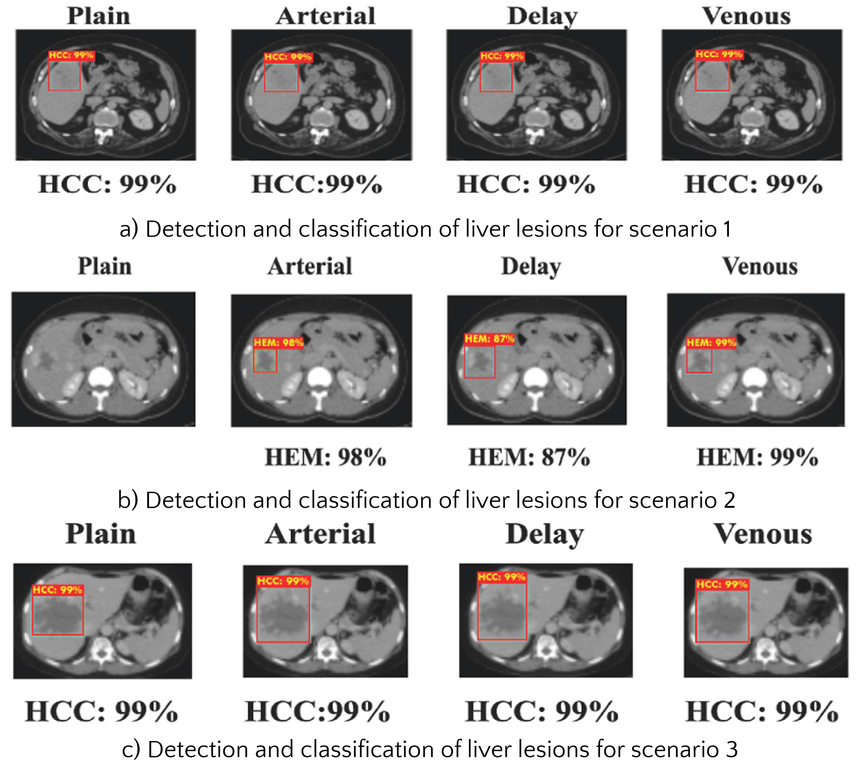
We conduct the training process on CT images before and after contrast injection for the detection and classification of liver lesions. After training, we proceed to the test phase.


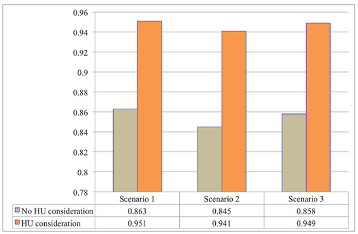
This indicates that scenario 1 is more efficient in extracting, detecting, and classifying liver lesions compared to scenarios 2 and 3.

Conclusions
In this work, we propose an efficient classification model for automatic detection and classification of liver lesions in CT images across four phases: plain, arterial, venous, and delay. The model leverages advanced deep learning networks (Faster R-CNN, R-FCN, and SSD) with various backbone networks like ResNet-101 and MobileNet-V2 for feature extraction. Incorporating HU attenuation in four-phase CT images enhances classification accuracy by aiding automatic segmentation and labeling of liver lesions. Experiments on multi-phase CT images of liver cysts, hemangiomas, and hepatocellular carcinoma reveal that considering HU density achieves 100% accuracy in lesion detection. Classification accuracy across three scenarios is 95.1%, 94.1%, and 94.9%, respectively, with scenario 1 performing best. The proposed methods outperform previous methods in accuracy, though the training time with large datasets remains a limitation. Future work will focus on optimizing training with parallel processing and expanding the study to include lesions in other body parts.
References
- Adam, A., Dixon, A.K., Gillard, J.H., Schaefer-Prokop, C., Grainger, R.G., Allison, D.J., 2014. Grainger & Allison's Diagnostic Radiology E-Book, sixth ed. Elsevier Health Sciences.
- Brant, W.E., Helms, C.A., 2012. Fundamentals of Diagnostic Radiology, fourth ed. Lippincott Williams & Wilkins.
- Dai, J., Li, Y., He, K., Sun, J., 2016. R-fcn: object detection via region-based fully convolutional networks. In: Advances in Neural Information Processing Systems, Barcelona, Spain, pp. 379–387. December 5-10.
- Girshick, R., Donahue, J., Darrell, T., Malik, J., 2014. Rich feature hierarchies for accurate object detection and semantic segmentation. In: Proceedings of the IEEE Conference on Computer Vision and Pattern Recognition, Columbus, OH, USA, pp. 580-587. June 23-28.
- Jang, H.J., Kim, T.K., Lim, H.K., Park, S.J., Sim, J.S., Kim, H.Y., Lee, J.H., 2003. Hepatic hemangioma: atypical appearances on ct, mr imaging, and sonography. Am. J. Roentgenol. 180, 135–141.
