Abstract
Brain haemorrhage is a critical problem with the high mortality rate that is typically diagnosed based on MRI/CT images. A lot of research is gaining attention recently for its high performance in image recognition of brain haemorrhage. In this paper, we propose a new approach, which can automatically detect, diagnose and classify of brain haemorrhages. Our proposed method focuses on analysing brain haemorrhage regions from images of brain haemorrhage. We rely on HU values to detect haemorrhage regions and determine the bleeding time of brain haemorrhages. It is useful for supporting doctors in timely treatment. Our experimental results show that the accuracy of detection of brain haemorrhages is 100% and the classification of brain haemorrhages achieves the accuracy of 93.33%.
Introduction
In recent years, there are many researchers in Vietnam and other countries in the world related to the medical field, particularly in detecting, diagnosing and classifying disease in humans. During treatment, subclinical results outcome is very important for doctors to detect, diagnose and treat, especially related to the brain haemorrhage. There have been many methods implemented to solve the problem, but there are still limitations. we propose a system that can automatically detect, diagnose and classify brain haemorrhages in patients from CT/MRI medical images. Our method implements a haemorrhaging segmentation based on Hounsfield Unit (HU) without performing image preprocessing. HU is computed easily and quickly from the values of Rescale Intercept and Rescale Slope available in medical image files of DICOM format. This is also a new contribution to our research.
In this work, we focus on the four types of brain haemorrhages namely epidural haematoma, subdural haematoma, subarachnoid haemorrhage and intracerebral haemorrhage. These considered haemorrhage types have many differences in aspects of visual features such as the size of the haemorrhage region, its shape, and its location within the skull... We also collect opinions from experienced doctors and specialists in the field of medical imaging in CanTho Medicine University Hospital to help us identify and distinguish the four popular types of brain haemorrhages from our datasets.

Decision Tree for High Blood Pressure Detection and Classifcation
Our method directly processes the source data on medical images formatted according to the Dicom standard without transforming into JPG, BMP, PNG, etc. This allows us to preserve valuable information for determining Hounsfield Units (HU) crucial for detecting brain hemorrhage and its timing. This approach represents a novel contribution to the field. We compute HU using a linear transformation based on a specific equation.
HU = Pixel_value * Rescale_slope + Rescale_intercept (1)
where: Pixel_value is the value of each image point, the Rescale_slope and Rescale_intercept are the parameter values provided in DICOM images.

Proposed Method
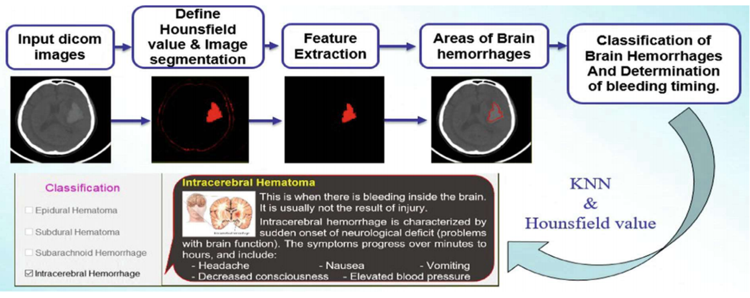
Our proposed method was described in Fig. 2 including the following stages:

- Stage 1: The determination of the HU value: the most valuable feature of the DICOM image data is ability to store a lot of the necessary information for the computation of the HU values.
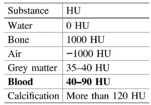
- Stage 2: Image segmentation: after computing the HU values of image points, we detect areas of brain hemorrhages by image segmentation based on HU values, which are in the range from 40 to 90.
- Stage 3: Determination of brain hemorrhage areas: we remove unrelated image areas and image areas due to some effects in CT technique such as the cortex.
- Stage 4: Feature extraction: we extract some important features from the areas of brain hemorrhages using the Regionprops tool in Matlab.
- Stage 5: Classification of brain hemorrhages: from extracted features, we apply the KNN algorithm using the Euclidean distance to identify brain hemorrhages.
- Stage 6: Determination of timing of haemorrhage: the bleeding timing was considered to support doctors making timely treatment for patients
Results
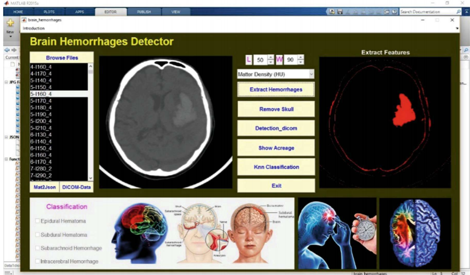
Our proposed method is experimented on a system that automatically detects, diagnoses, and classifies brain hemorrhages. We collected 500 CT images by DICOM standard from the patients' skull in CanTho University Hospital. The training and testing data sets are selected at a 7:3 ratio. It means that the training set of 350 CT images is classified into four types of brain hemorrhages based on the experience of doctors and specialists of Can Tho University Hospital. This data set includes 95 CT images of Epidural Hematoma, 85 CT images of Subdural Hematoma, 80 CT images of Subarachnoid Hemorrhage, and 90 CT images of Intracerebral Hemorrhage. The files of the training set are 179 Mb. The testing set of 150 images is classified into types of brain hemorrhages and normal brain (no bleeding), which includes 45 normal brain images, 25 images of Epidural Hematoma, 20 images of Subdural Hematoma, 30 images of Subarachnoid Hemorrhage, and 30 images of Intracerebral Hemorrhage.


From the confusion matrix shown in Table 2, the average accuracy is computed:

After identification and classification of the brain haemorrhages, our proposed method determines the bleeding time of brain to support doctors finding timely and effective treatments

Conclusions
There were various techniques developed for effectively detecting the haemorrhage in the brain. A method of automatically classifying brain haemorrhages is proposed in our research based on the Hounsfield Unit. Our proposed method identifies areas of brain haemorrhage better than some well-known image segmentation methods. It also improves the classification accuracy compared to these methods. In this paper, we present a truly automatic image segmentation method because it does not require a user to determine image-specific parameters such as thresholds or regions of interest. The use of the HU analysis leads to an important improvement in the determination of the regions and the bleeding timing of brain haemorrhages to support doctors making timely treatment. Our system can assist doctors in detecting and classifying the brain haemorrhages through medical images (CT/MRI) with DICOM standard, especially the bleeding timing. The four types of brain haemorrhages namely epidural haematoma, subdural haematoma, subarachnoid haemorrhage and intracerebral haemorrhage are considered in this paper.
References
- Ahmed, H.S.S., Nordin, M.J.: Improving diagnostic viewing of medical images using enhancement algorithms. J. Comput. Sci. 7(12), 1831-1838 (2011)
- Al-Ayyoub, M., Alawad, D., Al-Darabsah, K., Aljarrah, I.: Automatic detection and classification of brain hemorrhages. WSEAS Trans. Comput. 12(10) (2013)
- CT scan - Mayo Clinic (2016). https://www.mayoclinic.org/. Accessed 20
- Otsu, N.: A threshold selection method from gray-level histograms. Automatica 11(285- 296), 23-27 (1975)
- Johnson, S.: Stephen Johnson on Digital Photography. O'Reilly, Sebastopol (2006)
- Hounsfield, G.N.: Med. Phys. 7, 283-290 (1980). PubMed
- Hoa, P.N., Van Phuoc, L.: CT Chan thuong dau. Medical publishing House Ho Chi Minh city branch (2010)
- Josephson, C.B., White, P.M., Krishan, A., Al-Shahi Salman, R.: Computed tomography angiography or magnetic resonance angiography for detection of intracranial vascular malformations in patients with intracerebral haemorrhage. The Cochrane Library (2014) 9. Clunie, D., Cordonnier, E.: Digital Imaging and Communications in Medicine (DICOM) - Application/DICOM MIME Sub-type Registration (2014)
- Altman, N.S.: An introduction to kernel and nearest-neighbor nonparametric regression. Am. Stat. 46(3), 175-185 (1992)
- Mustra, M., Delac, K., Grgic, M.: Overview of the DICOM standard (PDF). In: 50th International Symposium ELMAR, Zadar, Croatia, pp. 39-44 (2008)
- Ferreira, A.J.M.: MATLAB Codes for Finite Element Analysis. Springer, Netherlands (2009). https://doi.org/10.1007/978-1-4020-9200-8
