Abstract
Kidney stones are the leading cause of illness and the incidence is increasing worldwide; untreated kidney stones can lead to complications such as urinary tract infections, kidney enlargement, and irreversible kidney failure; causing economic damage or even human lives if not treated promptly. Therefore, early detection and treatment of kidney stones are essential. Additionally, the application of deep learning networks has emerged as a competitive alternative to convolutional neural networks in the fields of computer vision, medical, and is widely used in various image recognition tasks. In this paper, we propose a method for detecting and segmenting kidney stones using state-of-the-art deep learning networks such as MobileNetV2, DenseNet169, NASNetMobile, and Vision Transformer (ViT); where YOLOV7 is integrated as a tool to automatically segment kidney stones, assisting doctors in quickly identifying the location of stones. The research results show that the proposed method achieves an accuracy of up to 99% on abdominal CT image datasets during training.
Introduction
Kidney stones are characterized by the formation of solid crystal masses in the urinary tract of the kidneys. There is significant variability in their pathophysiology, risk factors, clinical progression, and treatment methods. Factors contributing to kidney stone formation include genetics, metabolism, and environmental conditions. Kidney stones are now recognized as a systemic disease indicator and a predictor of metabolic and cardiovascular complications. They can cause pain, infection, urinary obstruction, kidney damage, and impact the patient's quality of life. In Vietnam, the incidence of kidney stones is on the rise. In the United States, approximately 1 in 1,000 adults are hospitalized annually for urinary stones, and about 1% of all autopsy cases reveal these stones. By age 70, up to 12% of men and 10% of women will have developed urinary stones, with sizes ranging from microcrystals to stones several centimeters in diameter. Coral stones, which can fill the entire renal pelvis, are particularly large. At age 70, 19.1% of men and 9.4% of women report having had kidney stones. The burden of this condition appears to be increasing, with the National Health and Nutrition Examination Survey recording a rise in self-reported kidney stone prevalence from 3.2% in 1976-1980 to 8.8% in 2014. This makes Vietnam one of the countries with the highest rates of kidney stones globally. Worldwide, the prevalence varies, with 1-5% in Asia, 5-9% in Europe, and 7-15% in North America. In Saudi Arabia, nearly 20% of the population suffers from kidney stones, while in China, the rate is only 4%. Iran, located in the stone belt, reports a prevalence of 4.2 per thousand. Global data indicate a rising prevalence of kidney stones in both genders over the last quarter of the 20th century, possibly due to environmental factors such as diet and lifestyle. However, the development of diagnostic processes for asymptomatic stones may partly explain this trend. Kidney stones increase with age and are more common in men than women.
Proposed Method
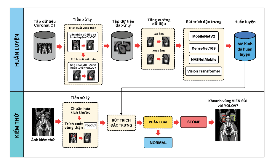
In this method, the detection of kidney stones uses the Vision Transformer model, consisting of two phases: training and testing, as illustrated in Figure 1.
Data Preprocessing:In the preprocessing phase, I label the kidney region data for the abdominal CT dataset.
Data Augmentation and Normalization: Next, I expand the proposed dataset by using the YOLOv7 model, which has been trained on the previous dataset, to extract kidney regions from Dataset 2. Additionally, I normalize the images to a size of 224x224 and perform data augmentation (Figure 6) by rotating the images by a maximum of 2% of the width or height and flipping the images horizontally.
Feature Extraction and Training:Based on the advantages discussed in section IIB, this phase involves feature extraction and training using the preprocessed dataset from the previous phase. The result of this phase is the trained models.

To detect and localize kidney stones, this study proposes several methods outlined in Table 1. Specifically, for Method 1, I train the YOLOv7 model on a labeled dataset to extract the kidney regions in CT images and simultaneously assist in localizing the kidney stones. Methods 2 through 4 are designed to provide diagnostic results, including either the presence of kidney stones or a normal condition.

Results
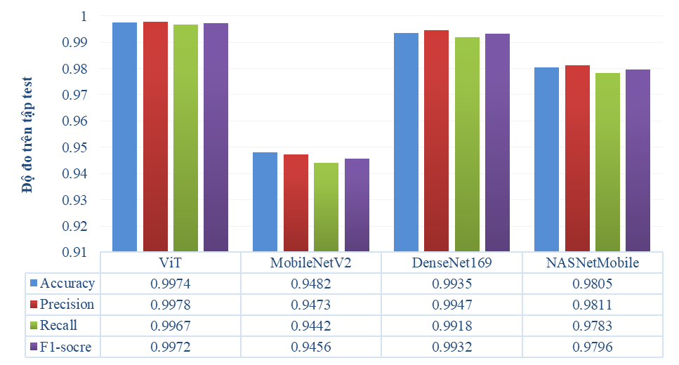
Figure 2 presents the performance metrics for the proposed scenarios. It can be concluded that the scenario utilizing the Vision Transformer architecture achieves higher results in detecting kidney stones compared to the advanced deep learning models proposed in the other scenarios

To evaluate the performance of the proposed network models, testing will be conducted across three scenarios: Stone (kidney stones present in both kidneys), Normal (no stones in either kidney), and Each of two (stones present in one of the two kidneys). The test results, including predictions and accuracy, are presented in Figure 3 through 5. Here, Stone refers to kidneys containing stones, while Normal refers to kidneys without stones (healthy).


Throughout the training and testing process, training parameters were adjusted to suit each proposed scenario. A comparison and evaluation were conducted, and the model evaluation criteria are summarized in Table 2.

Conclusions
Kidney stones are a major cause of disease with increasing prevalence worldwide, leading to economic damage or even loss of life if not treated promptly. This paper proposes a model for detecting and localizing kidney stones based on the Vision Transformer architecture, various deep learning networks, and YOLOv7 using a CT abdomen dataset. The YOLOv7 model demonstrated superior performance in object localization (stones within the kidneys). Additionally, we provide experimental results for various cases on CT images, including Normal (both kidneys healthy), Stone–Normal (one kidney with stones), and Stone (both kidneys with stones). The study results indicate that the proposed method achieves an accuracy of up to 99% on the CT abdomen image dataset during training.
References
- S. Shastri, J. Patel, K. K. Sambandam, and E. D. Lederer, “Kidney Stone Pathophysiology, Evaluation and Management: Core Curriculum 2023,” American Journal of Kidney Diseases, vol. 82, no. 5. 2023. doi: 10.1053/j.ajkd.2023.03.017.
- C. tịch H. T. N.-T. H. V. N. PGS.TS.BS.Vũ Lê Chuyên, “Tán sỏi thận qua da,” Trung tâm Kiểm soát bệnh tật TP. HCM.
- M. D. C. K. S. C. Glenn M. Preminger, “Sỏi tiết niệu,” https://www.msdmanuals.com/
- D. Aune, Y. Mahamat-Saleh, T. Norat, and E. Riboli, “Body fatness, diabetes, physical activity and risk of kidney stones: a systematic review and meta-analysis of cohort studies,” European Journal of Epidemiology, vol. 33, no. 11. 2018. doi: 10.1007/s10654-018-0426-4.
- R. H. Ma, X. B. Luo, Q. Li, and H. Q. Zhong, “Systemic analysis of urinary stones from the Northern, Eastern, Central, Southern and Southwest China by a multi-center study,” BMC Urol, vol. 18, no. 1, 2018, doi: 10.1186/s12894-018-0428-2.